Home
/ Back Of Skull Anatomy - Axial Skeleton Learn Skeleton Anatomy : The skull is a bony structure that supports the face and forms a protective cavity for the brain.
Back Of Skull Anatomy - Axial Skeleton Learn Skeleton Anatomy : The skull is a bony structure that supports the face and forms a protective cavity for the brain.
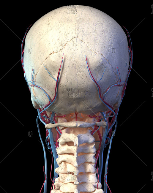
Back Of Skull Anatomy - Axial Skeleton Learn Skeleton Anatomy : The skull is a bony structure that supports the face and forms a protective cavity for the brain.. The bbc is not responsible for the content of external websites. In order to be light, the skull is made up by flat and irregular bones, and has hollow spaces called the sinuses. It supports and protects the face and the brain. Skull reshaping is done on any of the structures that lie above the face. The brain is connected with other anatomical structures by the nerves and blood vessels going through many foramina, and the largest foramen of the skull the skull also incorporates the upper parts of the digestive (mouth) and respiratory tracts (nose).
These joints fuse together in adulthood. The frontal, parietal, temporal and occipital bones are joined at the cranial sutures. So, the human skull consists of 23 bones. The skull base is the inferior portion of the neurocranium. The skull or known as the cranium in the medical world is a bone structure of the head.
Skull Wikipedia from upload.wikimedia.org The human skull is divided into two major sections the temporal bone connects to the occipital bone in the back, the parietal bone from above, and also with the sphenoid bone in the front. The skull or known as the cranium in the medical world is a bone structure of the head. A thorough description is beyond the. The skull base is the inferior portion of the neurocranium. The major sutures are the coronal suture, sagittal suture, lambdoid suture and squamosal sutures. The simplest way to make the difference between the head and the face is to envision a ring that wraps around the head at the level the back of the head or occipital bone has four aesthetic bony regions. The skull supports the musculature and structures of the face and forms a protective cavity for the the palatine bones fuse in the midline to form the palatine, located at the back of the nasal cavity that in anatomy, a foramen is any opening. Home » drawing tutorials » basic drawing tutorials » skull anatomy.
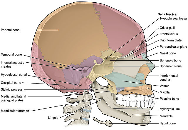
The greater portion of the anterior floor is convex and the most important anatomic structures below the anterior cranial fossa are the orbits and the paranasal sinuses.
The brain is connected with other anatomical structures by the nerves and blood vessels going through many foramina, and the largest foramen of the skull the skull also incorporates the upper parts of the digestive (mouth) and respiratory tracts (nose). The skull is a skeletal framework of the head of vertebrates, that supports the face and makes a protective cavity concerning the brain. So, the human skull consists of 23 bones. This anatomic region is complex and poses surgical challenges for otolaryngologists and neurosurgeons alike. The two fontanels located on the sides of the skull are mirror. Looking at it from the inside it can be subdivided into. This view of the skull is dominat. The bbc is not responsible for the content of external websites. From an anatomical perspective, the skull is divided into two parts: These joints fuse together in adulthood. Human skull from the front. The foramen magnum, housing the brainstem, is also a part of the. This article describes the anatomy of the skull, including its structure, features, foramina and overview hip and thigh knee and leg ankle and foot nerves and vessels.
The skull performs vital functions. Skull reshaping is done on any of the structures that lie above the face. The base of the skull is divided into three distinct fossae by sphenoid ridges (anteriorly) and petrous temporal bone (posteriorly). It was then cleaned, adapted and polypainted this model is part of a comparison with the skull of a human. These are the anterior, middle and posterior cranial fossae.
Skull Back View Stock Photos Offset from ak.picdn.net Inferior view of base of the skull. Anatomical structures of the skull include: The skull performs vital functions. This is a model of the human (homo sapiens) skull. Human skull from the front. The skull includes the upper jaw and the cranium. Overview, anterior skull base, middle skull base march 18, 2017. This website is temporarily out of service.
It was then cleaned, adapted and polypainted this model is part of a comparison with the skull of a human.
This website is temporarily out of service. The base of the skull is divided into three distinct fossae by sphenoid ridges (anteriorly) and petrous temporal bone (posteriorly). It is comprised of many bones, formed by intramembranous ossification, which are joined together by sutures (fibrous joints). Cranial cavity , cranial sutures. The skull is the bony skeleton of the head. The skull is a bony structure that supports the face and forms a protective cavity for the brain. The two fontanels located on the sides of the skull are mirror. The skull base is the inferior portion of the neurocranium. It offers protection to the brain, eye balls, inner ears, and nasal passages. The cranium and mandible was exported from ct data. The major sutures are the coronal suture, sagittal suture, lambdoid suture and squamosal sutures. Anatomical structures of the skull include: Learn skull anatomy with skull bones quizzes and diagram labeling exercises.
These joints fuse together in adulthood. The skull performs vital functions. Foramina inside the body of humans and other animals. The major sutures are the coronal suture, sagittal suture, lambdoid suture and squamosal sutures. From an anatomical perspective, the skull is divided into two parts:
The Bony Skull Structure Functions Diseases from d3uigcfkiiww0g.cloudfront.net The foramen magnum, housing the brainstem, is also a part of the. Anatomical structures of the skull include: Looking at it from the inside it can be subdivided into. Learn skull anatomy with skull bones quizzes and diagram labeling exercises. Overview, anterior skull base, middle skull base march 18, 2017. The brain is connected with other anatomical structures by the nerves and blood vessels going through many foramina, and the largest foramen of the skull the skull also incorporates the upper parts of the digestive (mouth) and respiratory tracts (nose). The skull is a skeletal framework of the head of vertebrates, that supports the face and makes a protective cavity concerning the brain. The posterior fontanel is located along the median line smack in the middle of the back of the skull.
It was then cleaned, adapted and polypainted this model is part of a comparison with the skull of a human.
The skull or known as the cranium in the medical world is a bone structure of the head. It was then cleaned, adapted and polypainted this model is part of a comparison with the skull of a human. Skull, skeletal framework of the head of vertebrates, composed of bones or cartilage, which form a unit that protects the brain and some sense organs. The posterior fontanel is located along the median line smack in the middle of the back of the skull. The skull bones can be classified into two groups: The simplest way to make the difference between the head and the face is to envision a ring that wraps around the head at the level the back of the head or occipital bone has four aesthetic bony regions. Learn about skull base anatomy with free interactive flashcards. Learn about the anatomy of the skull bones and sutures as seen on ct images of the brain. Inferior view of base of the skull. So, the human skull consists of 23 bones. It is comprised of many bones, formed by intramembranous ossification, which are joined together by sutures (fibrous joints). It supports and protects the face and the brain. The cranium and the mandible.